


I. 서 론
죽상동맥경화증(atherosclerosis)은 동맥 내에 죽상판 또는 섬유지방판을 형성하는 질환으로 혈관의 내강을 좁히고, 중막을 약화시켜 심근경색, 뇌경색 및 대동맥 동맥류 등의 합병증을 초래한다1.
죽상동맥경화증은 이전에는 일반적으로 퇴행성 과정으로 인식되었으나, 지금은 많은 종류의 세포들과 이들이 분비하는 성장인자, cytokine들이 얽혀서 동맥 경화증의 발생에 관여하는 것으로 밝혀졌다2.
이는 혈관 내피 염증 발생과 연관되며, 죽상동맥경화증의 발생 기전에 혈관내피의 염증 발생 단계는 중요하다. 혈관 내피의 염증은 산화적인 손상에 의해 유발되고, 염증 진행과정에서 단핵구가 혈관의 내피에 부착하는 과정이다3.
단핵구는 세포부착분자인 ICAM-1(Intercellular cell adhesionmolecule-1), VCAM-1(Vascular celladestion molecule-1)을 통해 혈관 내피세포에 부착하여 죽상동맥경화증의 진행에 영향을 미친다4.
현재는 죽상동맥경화증이 직접적으로 치료하기보다 죽상동맥경화증의 원인이 되는 혈전을 예방하는 목적으로 약물치료가 쓰이고 있다. 그러나 기존 약물을 통한 치료가 갖는 부작용으로 안전하고 효과적이며 직접적인 치료 효과가 있는 약물 개발이 필요한 실정이다5-7.
II. 재료 및 방법
본 실험에 사용한 갈근황련황금탕(Galkunhwanglyeon hwanggum-tang, 이하 GGT)과 소풍활혈탕(Sopunghwalhyeal -tang, 이하 SPT)의 구성약재는 대전한약국에서 구입 하였으며, 대전대학교 TBRC-RIC에 정선 후, 사용하였다. 본 실험에 사용한 처방의 내용 및 분량은 다음과 같다(Table 1, 2).
Table 1
The Prescription of GGT
Table 2
The Prescription of SPT
1. 시료 추출
GGT와 SPT를 구성하는 각각 약재들의 1첩 분량에 증류수를 500 ml씩 넣고 3시간 동안 환류추출한 후, 여과액을 rotary vacuum evaporator로 감압 농출하였으며, 농축된 용액을 freeze dryer로 동결 건조하였다. GGT는 14.27 g(수율 22.29%), SPT는 6.51 g(수율 13.85%)의 분말을 획득하였으며, 초저온(-80 °C) 냉동고에서 보관하여 실험에 필요한 농도로 증류수에 희석사용하였다.
2. 세포 배양
Human umbilical vein endothelial cell(HUVEC)은 0.2% gelatin solution으로 코팅된 plate에 EGMTM-2 medium과 EGMTM -2 singlequotsTM kit로 혼합된 배지를 사용하여 37 °C, 5% CO2 조건이 유지되는 세포배양기에서 배양하였으며, 2-3일 주기로 계대 배양하여 실험을 진행하였다.
3. 항염증효능 평가
1) 유전자 발현량 측정
(1) RNA 추출
6 well plate에 HUVEC을 1×10⁶ cell/well로 분주하여 24시간 동안 배양하였다. 배양 후 GGT, SPT를 각 50, 100, 200 μg/ml의 농도로 처리하고 2시간 뒤, 10 ng/ml의 TNF-α를 함께 처리하여 다시 6시간 동안 배양하였다. 이후, 1200 rpm에서 5분 동안 원심분리하여 얻은 세포에 easy blue 1 ml와 chloroform 200 μl를 넣고 vortexing 해준 후, 13000 rpm, 4 °C에서 10분 동안 원심분리하였다. 상층액 400 μl와 binding b uffer 400 μl를 실온에서 1분 동안 반응시킨 뒤 반응액 700 μl를 column에 주입하여 13000 rpm에서 30초 동안 원심분리하였다. Column에 washing buffer A를 700 μl 넣고 13000 rpm에서 30초 동안 원심 분리 후, washing buffer B를 700 μl 넣고 동일하게 원심분리하였다. Column 하단을 1.5 ml tube로 교체한 후, column에 elution buffer를 30 μl 넣고 1분 동안 반응시킨 뒤 13000 rpm에서 1분 동안 원심분리하여 total RNA를 추출하였다.
(2) cDNA 합성
역전사(reverse transcription) 반응은 RT premix kit의 mixture(reaction buffer, dNTPs mixture, RNase inhibiror, stabilizer, oligo dT15 primer), total RNA를 1 μg 넣고 DEPC-DW을 최종 부피가 20 μl가 되도록 첨가하였다. 이 혼합액을 잘 섞은 후, 45 °C에서 60분 반응시켜 first-strand cDNA를 합성하고, 95 °C에서 5분 동안 방치하여 M-MLV RT를 불활성화시켜 합성이 완료된 cDNA를 polymerase chain reaction(PCR)에 사용하였다.
(3) 유전자 증폭
합성이 완료된 cDNA를 증폭시키기 위하여 real-time PCR을 진행하였으며, real-time용 tube에 cDNA 1 μl, 각 primer 2 μl, SYBR Green 10 μl, DEPC-DW 5 μl 씩 넣어 95 °C에서 2분 동안 반응시키고 다음 95 °C에서 5초, 62.5 °C에서 30초를 40회 반복하여 전자를 증폭시켰다. 유전자 발현량은 대조군에 비하여 계산하였으며, 사용된 primer의 sequence는 Table 3과 같다.
Table 3
The Sequences of Primers
III. 실험결과
1. MCP-1
MCP-1 유전자 발현량을 측정한 결과, 대조군은 1.00±0.00 MCP-1/β-actin로 나타나 정상군에 비해 유의성 있는(+ : p<0.05, ++; p<0.01, +++ ; p <0.001) 발현량을 보였다. GGT와 SPT는 모든 농도에서 대조군에 비해 유의성 있는(* ; p <0.05, ** ; p 0.01, *** ; p<0.001) 감소가 나타났다(Table 4, Fig. 1).
Table 4
Effect of GGT and SPT on MCP-1 mRNA Expression Level in HUVEC
| Concentration (μg/ml) | Fold change (MCP-1/β-actin) | |
|---|---|---|
|
|
||
| GGT | SPT | |
| Normal | 0.04±0.01 | |
|
|
||
| Control | 1.00±0.00+++ | |
|
|
||
| 50 | 0.39±0.21* | 0.01±0.01*** |
| 100 | 0.19±0.13** | 0.00±0.01*** |
| 200 | 0.05±0.03*** | 0.01±0.01*** |
Fig. 1
Effect of GGT and SPT on MCP-1 mRNA expression level in HUVEC.
HUVEC were treated by 50, 100 and 200 μg/ml of GGT and SPT with 10 ng/ml TNF-α for 6 h. MCP-1 mRNA expression level were measured by polymerase chain reaction. The normal group were not treated TNF-α. The result were presented by the mean±S.D from three independent experiments (Significance of results, * ; p<0.05, ** ; p<0.01, *** ; p<0.001 compared to control).

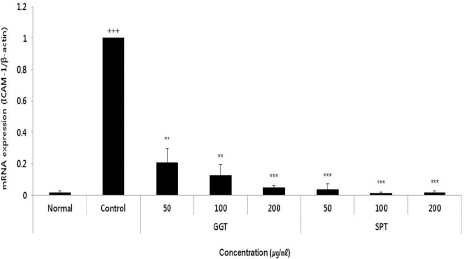
2. ICAM-1
ICAM-1 유전자 발현량을 측정한 결과, 대조군은 1.00±0.00 ICAM-1/β-actin로 나타나 정상군에 비해 유의성 있는(+ ; p<0.05, ++ ; p<0.01, +++ ; p<0.001) 발현량을 보였다. GGT와 SPT는 모든 농도에서 대조군에 비해 유의성 있는(** ; p<0.01, *** ; p<0.001) 감소가 나타났다(Table 5, Fig. 2).
Table 5
Effect of GGT and SPT on ICAM-1 mRNA Expression Level in HUVEC
| Concentration (μg/ml) | Fold change (ICAM-1/β-actin) | |
|---|---|---|
|
|
||
| GGT | SPT | |
| Normal | 0.02±0.01 | |
|
|
||
| Control | 1.00±0.00+++ | |
|
|
||
| 50 | 0.21±0.09** | 0.04±0.04*** |
| 100 | 0.13±0.07** | 0.01±0.01*** |
| 200 | 0.05±0.02*** | 0.02±0.01*** |
Fig. 2
Effect of GGT and SPT on ICAM-1 mRNA expression level in HUVEC.
HUVEC were treated by 50, 100 and 200 μg/ml of GGT and SPT with 10 ng/ml TNF-α for 6 h. ICAM-1 mRNA expression level were measured by polymerase chain reaction. The normal group were not treated TNF-α. The result were presented by the mean±S.D from three independent experiments (Significance of results, ** ; p<0.01, *** ; p<0.001 compared to control).
3. VCAM-1
VCAM-1 유전자 발현량을 측정한 결과, 대조군은 1.00±0.00 VCAM-1/β-actin로 나타나 정상군에 비해 유의성 있는(+ : p<0.05, ++ ; p<0.01, +++ ; p<0.001) 생성량을 보였다. GGT와 SPT는 모든 농도에서 대조군에 비해 유의성 있는(** ; p<0.01, *** ; p<0.001) 감소가 나타났다(Table 6, Fig. 3).
Table 6
Effect of GGT and SPT on VCAM-1 mRNA Expression Level in HUVEC
| Concentration (μg/ml) | Fold change (VCAM-1/β-actin) | |
|---|---|---|
|
|
||
| GGT | SPT | |
| Normal | 0.03±0.01 | |
|
|
||
| Control | 1.00±0.00+++ | |
|
|
||
| 50 | 0.12±0.07** | 0.02±0.02*** |
| 100 | 0.06±0.05*** | 0.03±0.02*** |
| 200 | 0.03±0.00*** | 0.03±0.01*** |
Fig. 3
Effect of GGT and SPT on VCAM-1 mRNA expression level in HUVEC.
HUVEC were treated by 50, 100 and 200 μg/ml of GGT and SPT with 10 ng/ml TNF-α for 6 h. VCAM-1 mRNA expression level were measured by polymerase chain reaction. The normal group were not treated TNF-α. The result were presented by the mean±S.D from three independent experiments (Significance of results, ** ; p<0.01, *** ; p<0.001 compared to control).

4. eNOS
eNOS 유전자 발현량을 측정한 결과, 대조군은 1.00±eNOS/β-actin로 나타나 정상군에 비해 유의성 있는(+: p<0.05, ++ ; p<0.01, +++ ; p<0.001) 발현량을 보였다. GGT는 모든 농도에서 대조군에 비해 유의성 있는(* : p<0.05, ** ; p<0.01) 증가가 나타났으며, SPT은 모든 농도에서 대조군에 비해 증가했으나 유의성은 나타나지 않았다(Table 7, Fig. 4).
Table 7
Effect of GGT and SPT on eNOS mRNA Expression Level in HUVEC
| Concentration (μg/ml) | Fold change (eNOS/β-actin) | |
|---|---|---|
|
|
||
| GGT | SPT | |
| Normal | 1.57±0.15 | |
|
|
||
| Control | 1.00±0.00+ | |
|
|
||
| 50 | 1.82±0.20* | 1.01±0.27 |
| 100 | 1.90±0.10** | 1.04±0.29 |
| 200 | 2.06±0.11** | 1.13±0.32 |
Fig. 4
Effect of GGT and SPT on eNOS mRNA expression level in HUVEC.
HUVEC were treated by 50, 100 and 200 μg/ml of GGT and SPT with 10 ng/ml TNF-α for 6 h. eNOS mRNA expression level were measured by polymerase chain reaction. The normal group were not treated TNF-α. The result were presented by the mean±S.D from three independent experiments (Significance of results, * ; p<0.05, ** ; p<0.01 compared to control).

IV. 고 찰
죽상동맥경화증은 심혈관질환의 위험인자인 나이, 혈압, 공복혈당, 콜레스테롤, 아포지방단백, 비만과 유의한 관련성을 보이며12 심혈관계 질환의 주된 요인으로 허혈성 심질환의 이환 및 사망13, 허혈성 뇌졸중 발생을 일으키는 주된 원인으로 알려져있다14.
2018년 발표한 통계청 보고에 따르면 심혈관계에 관련된 심장질환, 뇌혈관질환으로 인한 사망인구는 2007년 10만 명당 각각 44.1명, 60명이었으며 2017년 60.2명, 44.4명으로 증가, 감소세의 차이를 보였다. 순환 계통의 질환으로 인한 사망률은 2007년과 비교 하여 2017년 1.6% 증가하는 경향을 보였다15.
죽상동맥경화증의 생성 기전으로 산화적 스트레스와 혈관내피세포에서의 염증 유발 과정이 주요기전으로 인식되고 있으며16,11, 이는 혈액 중의 단핵구가 산화된 LDL에 의해 내막 안으로 유입되면서 시작된다. 단핵구는 대식세포로 분화되어 산화된 LDL을 탐식하고 활성화된 대식세포가 되어 거품세포를 형성하여 평활근 세포에 축적된다17. 활성화된 대식세포가 염증반응의 전사인자인 nuclearfactor κB(NFκB)를 촉진시키며, 여러 cytokine과 활성산소종을 유발시킨다. 그 결과 세포부착분자인 Intracellular adhesion molecule-1(ICAM-1), vascular cell-adhesion molecule-1(VCAM-1)과 monocyte chemoattractant protein-1(MCP-1) 등을 발생시켜 혈액 중의 단핵구 유입과 지속적인 염증반응들을 일으켜 죽상동맥경화증을 진행시킨다18-20.
일반적으로 약물치료는 동맥경화를 직접적으로 치료하기보다 혈소판제제, 혈전용해제, 항응고제 등으로 동맥경화의 원인이 되는 혈전성 질환을 예방하는 목적으로 쓰이고 있다. 그 중 항혈소판제제가 혈전성 질환을 예방하기 위해 보편적으로 쓰이고 있으나 위장장애, 출혈성 부작용 및 혈액학적 변화, 간독성, 과민성 반응 등의 부작용이 보고되고 있다. 그러므로 안전하고 효과적인 약물 개발에 대한 수요가 증가하고 있는 상태이다5-7.
葛根黃連黃芩湯은 ≪傷寒論≫21 <辨太陽病篇> 수록된 처방으로 급성장염, 역리, 소화불량 등으로 인한 下利, 또는 기관지천식 등에 사용되어왔다. 疎風活血湯은 沈의 ≪沈氏尊生書≫22에 수록된 처방으로 當歸, 川芎, 威靈仙, 白芷, 防己, 黃柏, 南星, 蒼朮, 姜活, 桂皮, 紅花 및 生薑으로 구성되어있으며, “或由風濕與痰, 如死血, 致走注刺痛, 其痛處或腫或紅, 則必宣邪通氣, 宜疎風活血湯”이라 기재된 이후, ≪東醫寶鑑≫23과 ≪證脈 方藥合編≫24에서 하면 이는 風濕痰과 瘀血로 인하여 四肢百節에 流注하여 刺痛하고 그 痛處가 或은 腫하고 或은 紅한 것을 치료한다 하였다.
다만 淸熱之劑로 염증을 억제하는 실험효과가 보고된 葛根黃蓮黃芩湯과 濕痰, 瘀血에 해당하는 동맥경화에 대한 실험효과가 검증된 疎風活血湯의 혈관염증억제효능을 비교 결과를 보고한 논문이 없어 그에 대한 의의가 있을 것으로 사료되었다. 이에 본 실험은 葛根黃蓮黃芩湯(GGT)와 疎風活血湯(SPT)의 한약리학적 효능에 착안하여 Human umbilical vein endothelial cel(HUVEC)에서 탕약의 추출물로 처리한 후 pro-inflammation이자 염증성 cytokine에 해당하는 TNF-a에 의해 유도된 CAMs (ICAM-1, VCAM1)와 동맥경화에 유관한 Chemotaxis에 해당하는 MCP-1 염증억제유발인자에 해당하는 e-NOS에 대한 GGT와 SPT의 효과를 비교하였다25-27.
사람의 혈관 내피세포에서 단핵구는 VCAM-1이나 ICAM-1과 같은 선택적으로 발현하는 다양한 세포부착분자를 통해서 결합한다. 세포부착분자는 단핵구의 부착을 중개하는데 이것은 특히 혈관에 많은 내피세포의 손상에서 흔하게 관찰된다. 단핵구가 세포부착분자에 의해 내피세포로 부착하는 것은 혈관염증의 초기단계이다18-20.
본 실험에서는 GGT와 SPT의 TNF-a로 유도된 단핵구의 세포부착이 GGT와 SPT 추출물에 의해 50, 100, 200 μg/ml 농도에 따라 유의하게 감소됨이 나타났다. 다만, 농도에 따라 유전자발현량에 있어서 SPT의 억제효과가 GGT의 억제 효과보다 우위성이 있음을 확인하였다.
Chemokine은 펩타이드 단백질의 일종으로 leukocyte의 주화성과 활성화에 영향을 끼친다. CC chemokine은 monocyte, lymphocyte, basophilic leukocyte 등을 유인한다28-30.
동맥경화 병변이 일어나기 쉬운 혈관 분지부 등에서 CC chemokine의 하나인 monocyte chemoattractant protein-1(MCP-1)의 발현이 증가하였으며31,32, 내피세포, macrophage, 평활근 세포에서 MCP-1의 mRNA의 발현이 확인되었다33. MCP-1은 동맥경화 진행과정에 있어서 subendothelial 세포층 속으로 monocyte의 침투를 유도한다34.
본 연구에서는 GGT와 SPT 추출물이 MCP-1 활성화에 어떠한 영향을 미치는 지에 대해 알아보았다. 그 결과, GGT과 SPT에서 50, 100, 200 μg/ml 농도에 따라 MCP-1의 유전자 발현량이 유의성 있게 감소되었다. 유전자 발현량에 있어서 SPT의 억제효과가 GGT에 비해 우위에 있음을 확인하였다.
이상 GGT와 SPT를 항염증에 대한 객관적 지표를 통한 실험적 결과를 살펴본 결과, GGT는 MCP-1과 같은 chemokine의 억제 효과에 우위에 있었다. GGT 는 MCP-1에 의해 유도된 단핵구 및 거품세포의 작용을 억제하여 평활근층 내 거품세포 침착을 억제하여 혈관염증 이전 단계에서 예방적 효과가 있을 것으로 기대된다. SPT는 ICAM-1, VCAM-1과 같은 CAMs의 생성을 억제하는 효과에 우위에 있었다. SPT는 염증 초기 단계 및 염증 진행과정을 억제하는 작용을 통한 동맥경화 예방 및 치료 효과가 있을 것으로 사료된다.


